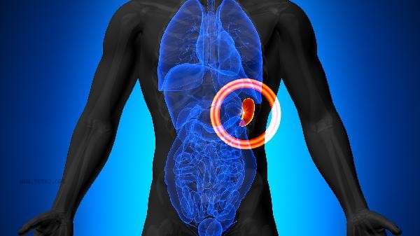
asymptomatic splenomegaly usually requires regular B-ultrasound examination to monitor changes in the spleen. Asymptomatic splenomegaly may be related to factors such as infection, blood system diseases, liver diseases, metabolic diseases, immune system diseases, etc. It is recommended to have a B-ultrasound examination every 3-6 months, combined with comprehensive evaluation of blood routine and liver function tests. Asymptomatic splenomegaly may not cause significant discomfort in the early stages, but continuous enlargement of the spleen increases the risk of rupture or suggests potential disease progression. B-ultrasound examination can clearly display the size, shape, and blood flow of the spleen. For patients with mild splenomegaly, a follow-up examination can be conducted every 6 months. If the spleen undergoes progressive enlargement or is accompanied by other abnormal indicators, a follow-up examination should be shortened to 3 months to further investigate the cause. Some patients with chronic liver disease may experience compensatory enlargement of the spleen due to portal hypertension, in which case liver stiffness and portal vein width need to be monitored simultaneously.
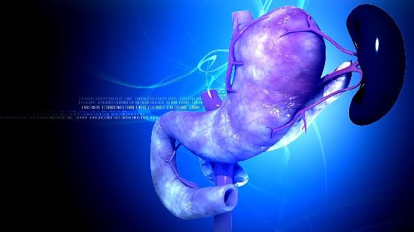
A few genetic metabolic diseases such as Gaucher's disease can also cause splenomegaly, and these patients need to undergo B-ultrasound every 3 months even if they are asymptomatic. For patients with spleen volume exceeding 50% of normal, it is recommended to undergo bone marrow aspiration or genetic testing to rule out hematological malignancies. Significant enlargement of the spleen may compress adjacent organs, leading to postprandial satiety or thrombocytopenia, and caution should be taken against the risk of spontaneous bleeding.
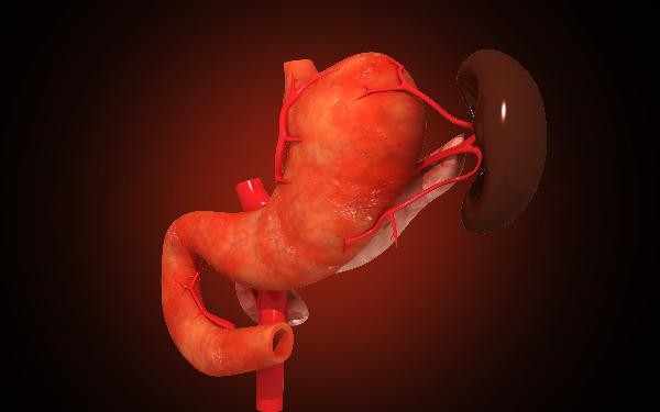
In daily life, one should avoid vigorous exercise and external force hitting the upper left abdomen, maintain a light diet, and abstain from alcohol. If a follow-up examination reveals rapid short-term enlargement of the spleen or symptoms such as fever and anemia, it is necessary to seek timely medical attention from a hematology or gastroenterology department. During regular follow-up, it is also necessary to pay attention to changes in platelet count and liver function indicators, and if necessary, further clarify the nature of splenic lesions through CT or MRI.


Comments (0)
Leave a Comment
No comments yet
Be the first to share your thoughts!