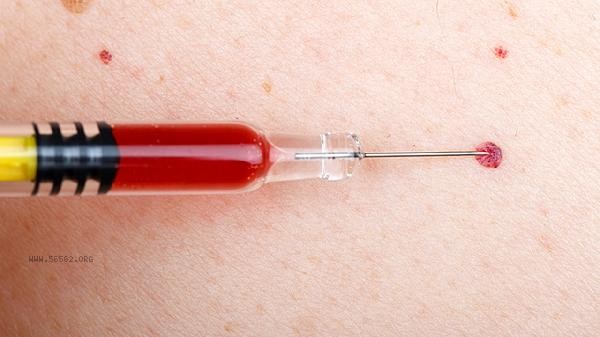
The bleeding time is usually shorter than the clotting time, mainly related to factors such as vascular constriction function, platelet activation rate, coagulation factor activation sequence, local tissue factor release, and fibrinolysis system regulation.

1. Vasoconstriction function:
Immediately triggers neuroreflex contraction after vascular injury, reducing blood loss. Endothelial cells release vasoconstrictors such as endothelin, causing the diameter of damaged blood vessels to shrink. This process is completed within seconds, earlier than the initiation of the coagulation cascade reaction.
2. Platelet activation rate:
Platelets adhere to damaged subendothelial collagen within 10 seconds and form temporary hemostatic plugs within 5 minutes. Platelets release substances such as ADP and thromboxane A2 to accelerate aggregation, which is 3-5 times faster than thrombin production.
3. Activation sequence of coagulation factors:

The tissue factor of exogenous coagulation pathway needs 6-8 seconds to be activated after exposure, and the endogenous pathway needs 1-2 minutes. The formation of platelet clots only takes 2-3 minutes, while the formation of complete fibrin clots takes 5-10 minutes.
4. Local tissue factor release:
Vascular outer membrane fibroblasts immediately release tissue factors, but they need to bind to factor VII to form complexes in order to activate downstream reactions. This process takes longer than platelet clot formation and has a shorter formation time.
3. Regulation of fibrinolytic system:
To prevent excessive coagulation, the fibrinolytic system begins to activate immediately after coagulation is initiated. Tissue type plasminogen activator continuously degrades fibrin, resulting in prolonged clotting time detection values, while bleeding time is not affected by this.

Hemostatic function can be maintained through daily supplementation of vitamin K such as spinach and broccoli, moderate exercise to enhance vascular elasticity, blood pressure control to reduce the risk of vascular damage, and other methods. The detection of bleeding time reflects the initial hemostasis efficiency, while the assessment of coagulation time evaluates the final coagulation stage. Combining the two can provide a more comprehensive assessment of hemostasis function. If abnormal elongation is found, it is recommended to complete specialized examinations such as vascular hemophilia factor testing and platelet function analysis. Maintaining a balanced diet and regular sleep schedule is crucial for maintaining normal coagulation mechanisms, and omega-3 fatty acids in deep-sea fish can help improve platelet function.


Comments (0)
Leave a Comment
No comments yet
Be the first to share your thoughts!