Early screening for lung cancer mainly relies on low-dose spiral CT examination, which is currently internationally recognized as the preferred method. Other auxiliary examinations include chest X-ray, tumor marker detection, sputum cytology examination, and bronchoscopy examination.
1. Low dose spiral CT:
Low dose spiral CT is the core method for early screening of lung cancer. Its radiation dose is much lower than conventional CT, but it can clearly display small lesions in the lungs. This examination can detect early lung cancer with a diameter of less than 1 centimeter, especially suitable for high-risk populations such as long-term smokers, those with a family history of lung cancer, or those with occupational exposure history. Through 3D reconstruction technology, doctors can accurately evaluate the morphology, density, and edge features of nodules, significantly improving the detection rate of early lung cancer. It is recommended that high-risk individuals undergo screening once a year. If suspicious nodules are found, follow-up observation or further examination should be conducted.
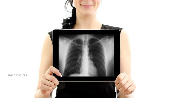
2. Chest X-ray:
Chest X-ray is a basic screening tool that has a certain display ability for larger tumors located in the hilum or mediastinum, but has low sensitivity for early small lesions. This examination has a low radiation dose and low cost, and is commonly used for preliminary assessment or health check ups. If abnormal shadows are found on X-rays, further low-dose CT or pathological examination is needed to clarify the nature. For asymptomatic low-risk populations, chest X-ray can be used as a routine screening option, but it cannot replace the core position of low-dose CT in early diagnosis.
3. Detection of tumor markers:
The detection of tumor markers helps to assess the risk of lung cancer through blood sampling and analysis of carcinoembryonic antigen, squamous cell carcinoma antigen, neuron specific enolase and other indicators. These markers may be mildly elevated in early lung cancer, but their specificity is limited, and some benign lung diseases may also cause numerical fluctuations. This examination cannot be used alone for diagnosis and needs to be combined with imaging results for comprehensive judgment. If the biomarker continues to rise or dynamically changes, it suggests the need for enhanced imaging follow-up. It is recommended that high-risk individuals undergo regular testing under the guidance of a doctor as a supplementary screening method.
4. Sputum cytology examination:
Sputum cytology examination analyzes the shed cells in sputum to search for cancer cells or atypical cells. This method has a high detection rate for central lung cancer such as squamous cell carcinoma, but lacks sensitivity for peripheral small lesions. The examination requires continuous collection of morning sputum, which is easy to operate and non-invasive, suitable for patients who cannot tolerate other examinations. If the result is positive, confirmation should be made through bronchoscopy or biopsy; If it is negative but highly suspected of lung cancer, it is still necessary to combine CT and other imaging methods. This examination is commonly used in high-risk populations with cough and sputum symptoms.
5. Bronchoscopy:
Bronchoscopy involves inserting a thin tube into the airway through the mouth or nose, directly observing bronchial mucosal lesions, and taking samples for biopsy. This examination has high diagnostic value for central lung cancer, as it can clarify the pathological type and gene mutation status. In early screening, if CT detects suspicious nodules near the airway, bronchoscopy can accurately obtain tissue specimens. Local anesthesia is required before examination, which may cause mild discomfort such as coughing or slight bleeding. For patients who are highly suspected of lung cancer but cannot be diagnosed by other examinations, bronchoscopy is an important diagnostic tool, but it is generally not used as a routine screening tool. Early screening for lung cancer should be based on individual risk factors, and low-dose spiral CT should be prioritized for high-risk populations, combined with auxiliary examinations such as tumor markers. Daily attention should be paid to quitting smoking, reducing exposure to air pollution, and seeking medical attention promptly if symptoms such as persistent cough and chest pain occur. After screening and discovering abnormalities, regular follow-up or further examination should be conducted according to medical advice, and early intervention should not be ignored due to lack of symptoms.


Comments (0)
Leave a Comment
No comments yet
Be the first to share your thoughts!