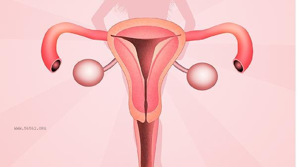
Vaginal endoscopy mainly examines lesions in the cervix, vagina, and external genitalia, especially precancerous lesions and early cervical cancer. It is an examination method that uses magnification technology to observe the mucosa of the lower reproductive tract, usually used to screen for abnormalities or diagnose suspicious lesions.

1. Cervical lesion screening:
Vaginal colposcopy is most commonly used to assess cervical health. When the screening results for cervical cancer are abnormal, such as cervical cytology examination indicating atypical cells or persistent infection with high-risk human papillomavirus, doctors will observe the morphology of cervical surface blood vessels and epithelium through colposcopy. During the examination, abnormal images such as acetic acid white epithelium, punctate blood vessels, or embedded blood vessels may be found. These manifestations are closely related to cervical intraepithelial neoplasia. Mild lesions such as CIN1 may self resolve, while CIN2 or CIN3 require further intervention. By using colposcopy to locate biopsy, the degree of the lesion can be determined, avoiding missed diagnosis of early cervical cancer.
2. Diagnosis of vaginal lesions:
Vaginal colposcopy is also used to detect abnormal hyperplasia or ulcers in the vagina. When patients experience contact bleeding, abnormal secretions, or rough vaginal walls, colposcopy can clearly display subtle changes in the vaginal mucosa. Common lesions include intraepithelial neoplasia of the vagina, which may appear as well-defined white patches or red erosion areas. Vaginal adenosis or vaginal endometriosis can also be identified through colposcopy, presenting as cystic structures or purple blue nodules. For postmenopausal women, mucosal fragility caused by vaginal atrophy also needs to be distinguished from cancer, and negative iodine test areas under colposcopy indicate the need for biopsy confirmation.
3. Assessment of external genital lesions:
Skin lesions in the external genital area are also within the scope of colposcopy examination. When there is itching, pigmentation, or ulcers in the external genitalia that do not heal for a long time, a colposcopy can magnify the distribution of blood vessels and the degree of keratinization in the external genitalia epithelium. Lichen sclerosus of the external genitalia presents as white atrophic patches, while intraepithelial neoplasia of the external genitalia may present as raised papules or wart like hyperplasia. During examination, attention should be paid to distinguishing between inflammatory changes and precancerous lesions, such as Paget's disease of the external genitalia, which often presents as well-defined red moist plaques. Vaginal colposcopy guided biopsy can improve diagnostic accuracy, especially for the detection of early vulvar cancer.
4. Follow up monitoring after treatment:
Patients who have undergone cervical conization or laser treatment should undergo regular evaluation of healing through colposcopy. After treatment, new blood vessels or scar tissue may appear on the wound, and colposcopy can distinguish between normal repair and residual lesions. For example, if the acetic acid white area persists during a follow-up examination 3-6 months after surgery, it may indicate the presence of residual lesions. For patients who have undergone cervical circular resection, colposcopy can also observe the narrowing or adhesion of the cervical canal. During long-term follow-up, the combination of colposcopy and human papillomavirus testing can more sensitively detect signs of recurrence and reduce the risk of cervical cancer.
5. Investigating the causes of abnormal bleeding:
Vaginal bleeding of unknown origin or bleeding after sexual intercourse can be traced using a colposcopy. Cervical polyps, submucosal fibroids, or cervical inflammatory hyperplasia appear as red protrusions under the microscope, with abundant surface blood vessels that are prone to bleeding. The mucosa of atrophic vaginitis is thin, congested, and prone to bleeding upon contact. In addition, colposcopy can detect small lesions hidden within the cervical canal, such as cervical polyps or early cervical cancer with cervical canal growth. For abnormal bleeding during pregnancy, vaginal colposcopy should be operated with caution, mainly to exclude bleeding caused by cervical polyps or cervical cancer, and avoid affecting the fetus. After vaginal colposcopy examination, the external genitalia should be kept clean, and sexual activity and baths should be avoided for 1-2 weeks to prevent wound infection. On the day of the examination, there may be a small amount of bloody discharge, which is a normal phenomenon. If the amount of bleeding after biopsy exceeds menstrual flow or is accompanied by fever, seek medical attention promptly. Daily attention to balanced nutrition and appropriate supplementation of vitamin C and folic acid can help repair cervical mucosa. Regular cervical cancer screening, including cytology and human papillomavirus testing, can provide a more comprehensive assessment of cervical health.


Comments (0)
Leave a Comment
No comments yet
Be the first to share your thoughts!