The deep vein, superior vena cava, and inferior vena cava belong to different anatomical structures. The deep vein is the venous system between the muscles of the limbs, while the superior vena cava and inferior vena cava are the main veins that collect blood from the upper and lower body and return to the heart, respectively.
1. Anatomical location:
Deep veins are distributed in the muscle spaces of the limbs, such as the brachial vein and ulnar radial vein in the upper limbs, and the femoral vein and popliteal vein in the lower limbs. The superior vena cava is located in the upper part of the chest cavity and is formed by the confluence of the left and right brachiocephalic veins; The inferior vena cava ascends along the right side of the spine and is formed by the confluence of the left and right common iliac veins.
2. Functional differences:
Deep veins mainly undertake the blood reflux of limb muscle tissue and have venous valves to prevent reflux. The superior vena cava is responsible for venous blood return to the head, neck, upper limbs, and chest, while the inferior vena cava collects venous blood from the abdomen, pelvic cavity, and lower limbs, both of which ultimately flow into the right atrium.
3. Clinical association:
Deep vein thrombosis is more common in the lower limbs and may cause pulmonary embolism. Upper vena cava obstruction can lead to facial and neck swelling, while inferior vena cava obstruction manifests as lower limb edema, abdominal wall varicose veins, and other signs.
4. Physical examination identification:
Ultrasound examination can clearly distinguish between deep vein and vena cava systems. Deep vein ultrasound focuses on observing valve function and thrombosis, while vena cava ultrasound needs to evaluate diameter, blood flow velocity, and adjacent organ compression.
5. Angiography:
interventional angiography shows segmental distribution of deep veins, Y-shaped structure of the superior vena cava, and long tubular structure of the inferior vena cava. CT or MRI three-dimensional reconstruction can display the anatomical relationship between the three in a three-dimensional manner.
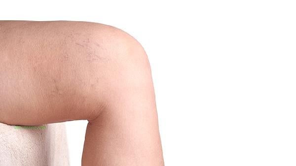
Avoid prolonged sitting and standing in daily life to prevent deep vein thrombosis, control blood pressure, and reduce endothelial damage. abnormalities in the vena cava are often associated with tumor compression, and it is recommended to undergo regular physical examinations including vascular ultrasound. Deep vein examination can choose lower limb vein ultrasound, while vena cava assessment requires chest and abdominal enhanced CT or magnetic resonance vein imaging. Wearing gradient pressure socks during exercise can help promote deep vein return. If there is unexplained limb swelling or chest and abdominal wall varicose veins, seek medical attention promptly.


Comments (0)
Leave a Comment
No comments yet
Be the first to share your thoughts!