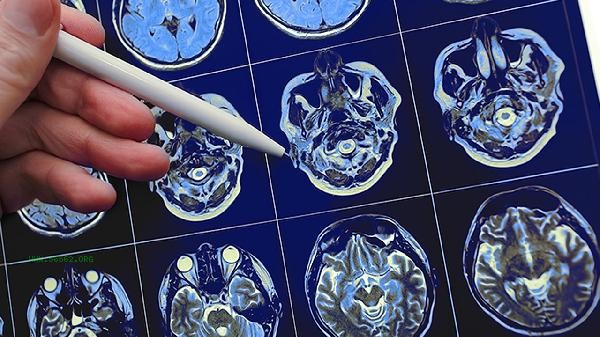
A decrease in heart rate to 50-60 beats per minute in patients with cerebral hemorrhage is a significant abnormality, and caution should be exercised regarding the risk of brainstem compression or increased intracranial pressure. Heart rate decline may be caused by factors such as vagus nerve stimulation, brain herniation, electrolyte imbalance, drug side effects, or secondary myocardial injury.

1. Vagus nerve stimulation: After cerebral hemorrhage, hematoma directly stimulates the medullary vagus nerve center, or the increase in intracranial pressure reflexively activates the vagus nerve, leading to a decrease in sinus node autonomy. At this time, there may be symptoms of parasympathetic nervous system excitation such as vomiting and pale complexion, and the bleeding site needs to be confirmed through head CT.
2. Brain Hernia Formation:
When the volume of intracranial hematoma increases, cerebellar tentorial hernia may compress the midbrain, causing early manifestations of Cushing's reaction. The typical triad is a decrease in heart rate, an increase in blood pressure, and irregular breathing, which is an emergency in neurosurgery and requires urgent dehydration and intracranial pressure reduction treatment.
3. Electrolyte imbalance:
Long term use of dehydrating agents may lead to hypokalemia, and when blood potassium is below 3.0 mmol/L, it can cause sinus bradycardia. Meanwhile, hypomagnesemia can also affect the myocardial conduction system, which needs to be confirmed through electrolyte testing. If necessary, intravenous supplementation of potassium magnesium aspartate is necessary.

4. Drug factors:
Some antihypertensive drugs such as metoprolol and diltiazem may excessively inhibit cardiac conduction, especially when combined with renal dysfunction, which increases the risk of drug accumulation. If the patient has recently used sedatives or mannitol, the medication plan needs to be re evaluated.
5. Myocardial injury:
Brain heart syndrome can cause stress-induced cardiomyopathy, with electrocardiogram showing QT interval prolongation and T wave inversion. Mild elevation of cardiac troponin indicates myocardial arrest caused by overactivation of the hypothalamic pituitary adrenal axis, requiring cardiac ultrasound evaluation of cardiac function. When patients with cerebral hemorrhage experience bradycardia, immediate electrocardiographic monitoring should be performed to maintain a head height of 30 degrees and avoid obstruction of intracranial venous return. Limit daily fluid intake to no more than 1500ml and prioritize high fiber foods to prevent intracranial pressure fluctuations caused by constipation. During the recovery period, passive limb movements can be performed to promote blood circulation, but drastic changes in posture should be avoided. It is recommended to monitor pupil changes and Glasgow Coma Scale every 2 hours. When the heart rate remains below 45 beats per minute or ventricular arrhythmia occurs, temporary pacemaker implantation should be considered.


Comments (0)
Leave a Comment
No comments yet
Be the first to share your thoughts!