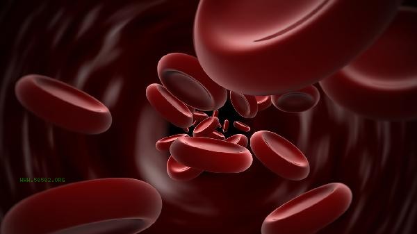
The main steps for using the red blood cell mean diameter detector include preparation before standardized operation, correct sample collection, equipment calibration and debugging, sample detection and analysis, and result recording and maintenance.

1. Preparation before operation
Before use, check whether the power connection is stable and confirm that the detector table is clean and dust-free. Take out the matching anticoagulant blood collection tube, check the expiration date and sealing. Wear sterile gloves and prepare consumables such as 75% alcohol swabs and disposable blood collection needles. After turning on the detector, it needs to be preheated for 5-10 minutes and can only be operated after the system self-test is completed.
2. Sample collection
adopts peripheral blood or venous blood collection method, and the blood collection site needs to be disinfected with alcohol swabs. The first drop of blood should be discarded during peripheral blood collection to avoid tissue fluid mixing and affecting the results. Blood samples and anticoagulants should be gently mixed 8-10 times immediately to prevent coagulation or hemolysis. The sample should be tested within 4 hours after collection to avoid changes in cell morphology.
3. Equipment Calibration
Before the first daily use, a standard calibration sample should be used for quality control to observe whether the absorbance value is within the allowable error range. If the deviation exceeds ± 5%, it is necessary to perform photoelectric calibration or mechanical zeroing according to the instructions. Regularly use particle size standard microspheres to calibrate the optical system, ensuring that the detection channel is unobstructed or the optical path is not offset.

4. Detection and analysis
Inject the mixed anticoagulant into a dedicated counting plate to avoid the generation of bubbles. Select the red blood cell diameter detection mode, set the sample number and detection parameters. After starting the detection, the instrument will automatically complete cell image acquisition, edge recognition, and diameter calculation. Abnormal results need to be retested 2-3 times to eliminate operational errors or sample interference factors.
5. Result Processing
Print or export the data report promptly after the detection is completed, recording the sample number and detection time. Rinse the counting plate and sample channel with physiological saline to prevent protein deposition. Before turning off the power, it is necessary to perform system maintenance procedures, regularly replace filters, and clean optical components. All quality control data should be archived for future reference. In case of persistent abnormalities, engineers should be contacted for maintenance. When operating the red blood cell mean diameter detector, it is necessary to strictly follow the standardized process, pay attention to the principle of sterility during the blood collection process, and avoid cell deformation caused by compression. The testing environment should maintain a temperature of 18-25 ℃ and a humidity of 40-60% to prevent the instrument from being affected by moisture or static interference. Regularly participate in inter room quality evaluations to compare the consistency of testing results from different equipment. Medical staff need to receive professional training, proficiently master the skills of cell morphology interpretation, and promptly review blood smears if abnormal red blood cell distribution curves are found.


Comments (0)
Leave a Comment
No comments yet
Be the first to share your thoughts!