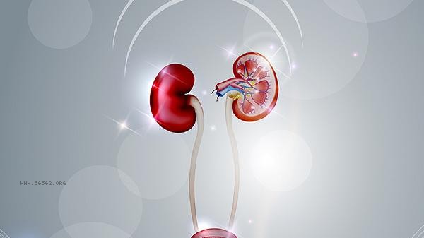
The examination of kidney function is mainly carried out through blood tests, urine tests, and imaging examinations. Common examination items include blood creatinine, blood urea nitrogen, estimated glomerular filtration rate, urine routine, and renal ultrasound.

1. Blood test:
Blood test is one of the most essential methods for evaluating kidney function. The main indicators include blood creatinine and blood urea nitrogen. When the renal filtration function decreases, these two substances accumulate in the blood, leading to an increase in their values. Doctors will also calculate the estimated glomerular filtration rate based on blood creatinine levels combined with factors such as the patient's age and gender, which can more accurately reflect the efficiency of the kidneys. In addition, cystatin C is also a sensitive indicator that is not affected by muscle mass and can detect mild renal function decline earlier. Before conducting a blood test, it is usually necessary to fast for 8-12 hours to avoid the impact of a high protein diet on the results.
2. Urine examination:
Urine examination is an important means of detecting early kidney damage. Urinalysis can detect abnormal components such as protein, red blood cells, white blood cells, or casts in urine. Positive urine protein usually indicates damage to the glomerular filtration membrane and is a sensitive signal in the early stages of kidney disease. Urinary microalbumin excretion rate is a key index for early screening of diabetes nephropathy and hypertensive nephropathy. When collecting urine samples, it is recommended to collect the first mid morning urine. Women should avoid their menstrual period to avoid interfering with the results.
3. Imaging examination:
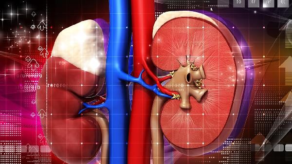
Imaging examination mainly observes whether there are any abnormalities in the morphology and structure of the kidneys. Renal ultrasound is a commonly used and non-invasive examination method that can clearly display the size, shape, and cortical thickness of the kidney, and determine whether there are structural problems such as renal cysts, kidney stones, hydronephrosis, or tumors. For situations that require a more detailed assessment of vascular or tumor lesions, doctors may recommend CT or magnetic resonance imaging examinations. These examinations do not require special preparation, but CT enhanced scans require the use of contrast agents, and patients with severe renal impairment need to carefully assess the risk.
4. Renal function imaging:
Renal function imaging is a nuclear medicine examination method that uses intravenous injection of radioactive tracers and special equipment to dynamically observe the uptake, distribution, and excretion of tracers in the kidneys, in order to evaluate the independent functions of the left and right kidneys separately. This examination has unique value in determining the blood supply, tubular function, and urinary tract obstruction of a unilateral kidney, especially for patients who require kidney surgery or evaluation of renal artery stenosis.
5. Renal biopsy:
Renal biopsy is the gold standard for obtaining renal pathological diagnosis, but it is an invasive examination. Under ultrasound guidance, doctors use a puncture needle to take a small amount of kidney tissue for pathological analysis, which can clarify the pathological type of glomerular disease, determine the degree of renal interstitial fibrosis, and evaluate the activity and reversibility of the disease. This examination is usually used for the diagnosis of unexplained acute kidney injury, massive proteinuria or nephrotic syndrome, as well as systemic diseases such as systemic lupus erythematosus involving the kidneys. After surgery, it is necessary to rest in bed for 24 hours and closely observe for complications such as hematuria or perirenal hematoma.
Daily protection of the kidneys should avoid the abuse of painkillers and herbal medicines with unknown ingredients, control blood pressure and blood sugar, maintain moderate water intake without holding urine, and undergo a urine routine and blood creatinine test once a year. In case of eyelid or lower limb edema, increased urine foam that is difficult to dissipate, abnormal urine color, such as meat washing water sample or significantly increased nocturnal urine frequency, etc., the doctor should go to the nephrology department in time to select the most appropriate examination combination according to the specific symptoms. Please avoid vigorous exercise before the examination. On the day of the examination, eat normally but do not drink a large amount of water to avoid diluting urine and affecting the results.


Comments (0)
Leave a Comment
No comments yet
Be the first to share your thoughts!