Checking for cerebral blood supply deficiency is mainly determined through a comprehensive evaluation of clinical symptoms, physical examination, and imaging examinations. Common methods include transcranial Doppler ultrasound, carotid artery ultrasound, head magnetic resonance imaging, head computed tomography angiography, and digital subtraction angiography.
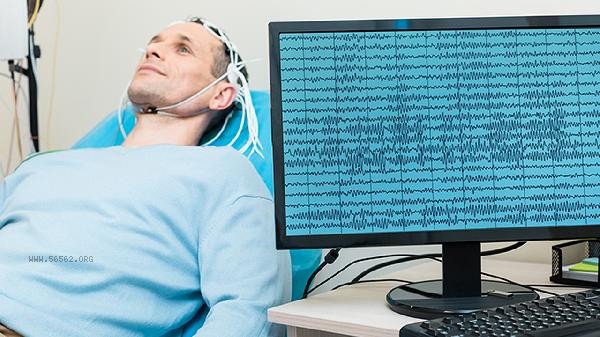
1. Transcranial Doppler Ultrasound:
Transcranial Doppler ultrasound is a non-invasive examination that uses ultrasound to detect the blood flow velocity, direction, and spectral morphology of major intracranial arteries, and can directly evaluate the hemodynamic status of blood vessels such as the middle cerebral artery, anterior cerebral artery, and posterior cerebral artery. If there is a decrease in blood flow velocity, an increase in vascular resistance, or abnormal spectral morphology, it may indicate insufficient cerebral blood supply. This examination has a good indication of vascular spasm, stenosis, or occlusion, but cannot directly display the structure of the vascular wall. It needs to be combined with other examinations for comprehensive judgment.
2. Carotid artery ultrasound:
Carotid artery ultrasound mainly examines the blood vessels supplying the brain to the neck, including the common carotid artery, internal carotid artery, and vertebral artery. It can clearly display whether there is plaque formation on the inner wall of the blood vessel, whether the lumen is narrow or occluded, and whether the blood flow velocity is abnormal. The carotid artery is the main pathway for blood supply to the brain. If there is severe stenosis or plaque shedding in this area, it can easily lead to insufficient blood supply to the brain or even cerebral infarction. This examination has no radiation and good reproducibility, and is commonly used for preliminary screening and long-term follow-up.
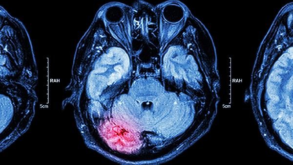
3. Head magnetic resonance imaging:
Head magnetic resonance imaging can provide detailed images of brain structures, including brain parenchyma, ventricles, blood vessels, etc. For cerebral ischemia, magnetic resonance imaging can display changes such as white matter lesions, lacunar infarction, or brain atrophy caused by long-term chronic ischemia. In addition, magnetic resonance angiography can non invasively display the morphology of major intracranial arteries and detect vascular stenosis, occlusion, or malformation. This examination has high resolution for soft tissue, but is not suitable for individuals with metal implants or claustrophobia in the body.
4. Head computed tomography angiography:
Head computed tomography angiography scans through intravenous injection of contrast agents, which can reconstruct intracranial and cervical blood vessels in three dimensions, clearly displaying the degree of vascular stenosis, plaque properties, and vascular wall calcification. This examination is relatively accurate in locating and quantitatively evaluating vascular lesions, especially suitable for rapid assessment of acute ischemic cerebrovascular events. But the examination requires the use of iodine containing contrast agents, and those with renal insufficiency or iodine allergy need to inform the doctor in advance.
5. Digital subtraction angiography:
Digital subtraction angiography is the "gold standard" for diagnosing cerebrovascular diseases. By injecting contrast agent into the carotid or vertebral artery through a catheter, it dynamically displays the morphology, blood flow velocity, and collateral circulation of cerebral blood vessels in real time. It can accurately evaluate lesions such as vascular stenosis, occlusion, aneurysms, or vascular malformations, providing direct basis for interventional treatment. But this examination is an invasive procedure with a certain risk of radiation exposure and bleeding at the puncture site, and is usually used when other non-invasive examinations cannot provide a clear diagnosis. If symptoms such as repeated dizziness, headache, memory loss, lack of concentration, or transient limb numbness and weakness occur, it is recommended to seek medical attention in a timely manner, and a neurologist should choose appropriate examination items based on the specific situation. Pay attention to controlling blood pressure, blood sugar, and blood lipids in daily life, quit smoking and limit alcohol consumption, maintain a regular schedule and moderate exercise, which can help prevent the occurrence of cerebral ischemia.


Comments (0)
Leave a Comment
No comments yet
Be the first to share your thoughts!