onychomycosis, commonly known as onychomycosis, can be diagnosed through physical examination, fungal microscopy, fungal culture, dermatoscopy, histopathological examination, and other methods. Onychomycosis is usually caused by fungal infections such as dermatophytes. It is recommended to seek medical attention promptly for a clear diagnosis.
1. Physical examination:
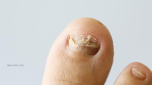
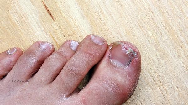
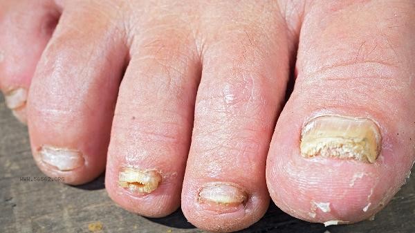
The doctor will observe the shape, color, texture, etc. of the nails with the naked eye. The nails of onychomycosis are usually characterized by turbidity, thickening, uneven surface, discoloration such as gray white or yellow brown, and separation of the deck from the nail bed. The doctor will also check for debris accumulation. This examination is a basic judgment, but cannot confirm the diagnosis. It needs to be combined with other examinations.
2. Fungal microscopy:
This is the most commonly used and rapid method for diagnosing onychomycosis. The doctor will scrape off the debris or deck fragments under the affected nail, place them on a glass slide, add potassium hydroxide solution to dissolve the keratin, and directly search for fungal hyphae or spores under a microscope. If the microscopic examination result is positive, it can be diagnosed as onychomycosis. However, there is a possibility of false negatives in this method, which means that sometimes a small number of fungi may not be detected.
3. Fungal culture:
When the fungal microscopy result is negative but highly suspected of onychomycosis, or when specific bacterial strains need to be identified to guide medication, this examination will be performed. Inoculate the collected specimens onto a culture medium and incubate at a suitable temperature for several weeks to observe for fungal growth. Positive cultivation can confirm the diagnosis and identify which fungus it is, such as Trichophyton rubrum, Trichophyton rubrum, etc.
4. Skin examination:
Skin examination is a magnifying glass with a light source that can non invasively observe the fine structure of nails. Under dermatoscopy, onychomycosis often presents characteristic changes such as serrated edges, longitudinal stripes, and accumulation of debris under the deck. This examination helps to distinguish onychomycosis from other nail diseases such as psoriasis and nail trauma, and can guide doctors in selecting the sampling site to improve the positive rate of fungal microscopy examination.
5. Histopathological examination:
For cases with difficult diagnosis, poor treatment effect, or suspected other lesions, this examination can be performed. The doctor will take a small piece of diseased tissue, fix it, slice it, stain it, and observe it under a microscope. If fungal hyphae or spores are found in the deck or nail bed tissue, the diagnosis can be confirmed. This is one of the gold standards for diagnosis, but it belongs to invasive examination and is less commonly used. After being diagnosed with onychomycosis, one should actively cooperate with doctors for treatment. Pay attention to keeping hands and feet clean and dry in daily life, and avoid sharing personal items such as slippers, towels, and nail clippers with others to prevent cross infection. Wear loose and breathable shoes and socks to avoid keeping nails in a damp environment for a long time. Patience is required during the treatment period, as nail growth is slow and usually takes several months to see significant improvement. Meanwhile, actively treating fungal infections in other parts of the body such as tinea pedis and tinea manus can help prevent the recurrence of onychomycosis.


Comments (0)
Leave a Comment
No comments yet
Be the first to share your thoughts!