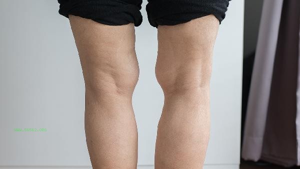
Muscle contracture usually requires a comprehensive diagnosis through electromyography, muscle biopsy, blood tests, imaging examinations, and nerve conduction tests. Muscle contraction may be related to neurological abnormalities, muscle diseases, metabolic disorders, trauma, or genetic factors. The specific examination plan should be developed by the doctor based on the symptoms.

1. Electromyography
Electromyography can distinguish between neurogenic or myogenic contractures by recording muscle electrical activity to determine the functional status of the neuromuscular system. During the examination, insert a fine needle electrode into the target muscle and observe the changes in electrical signals during rest and contraction. Peripheral neuropathy, motor neuron disease, etc. can lead to abnormal spontaneous potentials, while muscular dystrophy is characterized by a shortened duration of motor unit potentials.
2. Muscle biopsy
Muscle biopsy is used to obtain muscle tissue for pathological analysis, and is suitable for diagnosing organic lesions such as myositis and metabolic myopathy. The sampling site is often selected from the biceps brachii or quadriceps femoris, and characteristic changes such as muscle fiber necrosis and inflammatory infiltration can be observed through special staining. Attention should be paid to the possibility of local hematoma or infection risk after biopsy.
3. Blood tests
Blood tests include screening for indicators such as creatine kinase, electrolytes, and thyroid function. Elevated creatine kinase indicates muscle damage, hypokalemia can cause periodic paralysis, and thyroid dysfunction may lead to muscle rigidity. For pediatric patients, amino acid and organic acid metabolism related indicators need to be tested to exclude genetic metabolic diseases.

4. Imaging examination
Ultrasound or magnetic resonance imaging can visually display muscle structural abnormalities, such as muscle fibrosis, hematoma, or space occupying lesions. Magnetic resonance imaging has high resolution for soft tissue and can evaluate the degree of muscle edema and fat infiltration. Ultrasound examination can dynamically observe the state of muscle contraction and assist in determining the mechanical factors of contraction.
5. Nerve conduction examination
Determination of nerve conduction velocity can evaluate peripheral nerve function and differentiate nerve compression or demyelinating lesions. By stimulating nerves with electrodes and recording muscle responses, a decrease in conduction velocity indicates myelin damage, while a decrease in wave amplitude may indicate axonal injury. This examination has high diagnostic value for compressive neuropathy such as carpal tunnel syndrome. After being diagnosed with muscle contracture, targeted treatment should be carried out based on the cause. Neurogenic contracture may require muscle relaxants such as diclofenac, while myogenic problems require nutritional support and rehabilitation training. It is necessary to maintain moderate muscle stretching in daily life, avoid prolonged fixed posture, and supplement magnesium and vitamin D to help maintain neuromuscular function. During acute attacks, hot compress can be used to relieve spasms. If recurrent or accompanied by muscle weakness symptoms, timely follow-up should be sought.


Comments (0)
Leave a Comment
No comments yet
Be the first to share your thoughts!