Targeted drug induced interstitial pneumonia can be treated through drug discontinuation observation, glucocorticoid therapy, oxygen therapy support, anti fibrotic drug intervention, and regular imaging monitoring. This complication is usually induced by drug toxicity, immune-mediated injury, abnormal repair of alveolar epithelial cells, genetic susceptibility, and co infection.
1. Drug discontinuation observation:
The immediate suspension of the use of relevant targeted drugs is the primary measure. Clinical data shows that about 30% of patients experience spontaneous relief of symptoms after discontinuation of medication, and close monitoring of changes in blood oxygen saturation and respiratory rate is necessary. During discontinuation, it is recommended to have a chest CT scan every 48 hours to assess the extent of lung infiltration.
2. Corticosteroid therapy:
Moderate to severe cases require methylprednisolone shock therapy, with an initial dose of 1-2mg per kilogram of body weight, gradually decreasing after symptom control. During hormone therapy, blood glucose, bone density, and infection indicators need to be monitored, with an average duration of 8-12 weeks.
3. Oxygen therapy support:

When the arterial oxygen partial pressure is below 60mmHg, oxygen therapy should be initiated. Using nasal high flow humidified oxygen therapy can improve alveolar ventilation function. Patients with severe respiratory failure should consider non-invasive ventilation support, and an oxygenation index less than 150mmHg is a critical indicator for tracheal intubation.
4. Anti fibrotic drugs:
For patients with progressive pulmonary fibrosis, pirfenidone or nintedanib can be used in combination. These drugs delay fibrosis by inhibiting the transforming growth factor beta pathway, and regular checks of liver function and weight changes are required during medication.
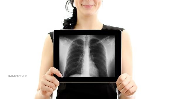
5. Imaging monitoring: During
treatment, high-resolution CT scans are performed every 2 weeks, with a focus on observing the extent of ground glass opacities and the degree of bronchial dilation. The 6-minute walk test and lung function DLCO testing should be conducted simultaneously, and the recovery of lung diffusion function often lags behind imaging improvement by 3-6 months.

During the rehabilitation period, patients should maintain moderate aerobic exercise such as swimming, Tai Chi, etc. to enhance lung function. The diet should choose high protein, vitamin E rich deep-sea fish and nut foods. The living environment should be controlled with humidity between 40% -60% to avoid exposure to allergens such as dust mites. It is recommended to re-examine lung function every 3 months and continue to track for more than 2 years. Before using targeted drugs again, bronchoalveolar lavage fluid testing should be performed to assess the risk of recurrence. If symptoms of shortness of breath or dry cough worsen after activity, timely follow-up should be sought.


Comments (0)
Leave a Comment
No comments yet
Be the first to share your thoughts!