Multiple cerebral small vessel lesions in the brain are mostly mild to moderate health problems, and their severity mainly depends on the extent of the lesions, accompanying symptoms, and the control of underlying diseases. The common influencing factors are aging, poor control of hypertension, complications of diabetes, hyperlipidemia and smoking history.
1. Age factor:
The detection rate of cerebral small vessel disease in people over 60 years old can reach 30% -50%, which is a common manifestation of cerebral vascular aging. The decrease in vascular wall elasticity leads to microarterial sclerosis, which usually only manifests as cerebral white matter rarefaction or lacunar infarction, and most do not have acute neurological deficit symptoms.
2. Effects of hypertension:
Long term uncontrolled hypertension can accelerate small artery hyaline degeneration, leading to multiple punctate ischemic lesions in subcortical white matter. These patients may experience mild cognitive decline or gait abnormalities, and the progression of the disease can be slowed down after reaching blood pressure standards.
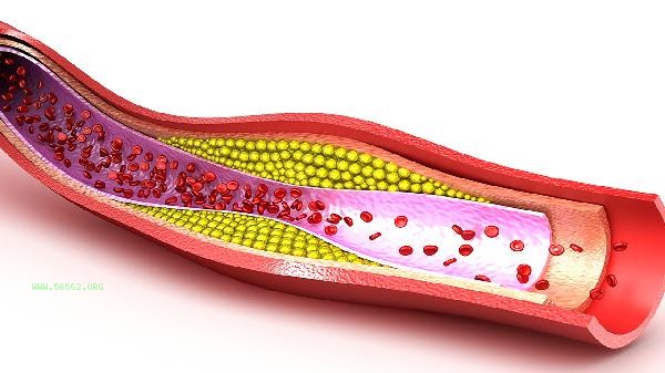
3. Metabolic syndrome:

diabetes and hyperlipidemia will cause vascular endothelial damage and form diffuse small vascular disease. The typical imaging manifestation is multiple lacunar lesions in the basal ganglia, which may be accompanied by transient ischemic attacks, but rarely cause extensive cerebral infarction.
4. Vasculitic changes:
A few immune diseases such as systemic lupus erythematosus may cause small vessel inflammatory lesions, which need to be diagnosed by combining anti nuclear antibody spectrum and other tests. Typical symptoms include recurrent headaches, cognitive impairment combined with other systemic symptoms.
5. Hemorrhagic transformation:
About 5% -10% of patients may experience microbleeds, which are common in people who take anticoagulant drugs for a long time. Magnetic susceptibility weighted imaging usually shows asymptomatic punctate bleeding, but the necessity of anticoagulant therapy needs to be evaluated.

It is recommended that patients undergo a follow-up cranial MRI every 6-12 months, with a focus on monitoring changes in the Fazekas grading of white matter lesions. It is recommended to maintain a low salt and low-fat diet in daily life. The Mediterranean dietary pattern is recommended, with a daily intake of over 300 grams of dark vegetables. Regularly engage in aerobic exercise such as brisk walking and swimming, accumulating 150 minutes per week. Strictly control the blood pressure below 140/90mmHg, and maintain the glycosylated hemoglobin of diabetes patients below 7%. Sudden dizziness, unclear speech, or physical weakness require immediate medical attention.


Comments (0)
Leave a Comment
No comments yet
Be the first to share your thoughts!