The vascular resistance index (RI) is calculated from the blood flow spectrum during ultrasound Doppler examination, and the formula is systolic peak flow velocity - end diastolic flow velocity/systolic peak flow velocity, with a normal range of 0.55-0.75. Abnormal RI values may reflect changes in vascular elasticity, hemodynamic changes, or organ perfusion abnormalities.
1. Ultrasonic detection principle:
RI calculation relies on pulse Doppler technology. The probe emits ultrasonic waves to receive red blood cell reflection signals, and converts blood flow signals into spectral waveforms through fast Fourier transform. During the examination, the straight segment of the target blood vessel should be selected, and the sampling volume should be adjusted to 1/3-1/2 of the vessel diameter. The angle between the sound beam and the blood flow should be ≤ 60 ° to ensure data accuracy.
2. Blood flow parameter measurement:
Identify the highest systolic point PSV and the lowest diastolic point EDV on the spectrogram, and manually or automatically measure the values of both. PSV reflects the maximum perfusion pressure of blood vessels, while EDV reflects the distal microcirculation resistance. The difference between the two, PSV-EDV, and the ratio of PSV to PSV, is the RI value. The measurement requires continuous collection of 3-5 cardiac cycles and taking the average value.
3. Clinical significance interpretation: An increase in

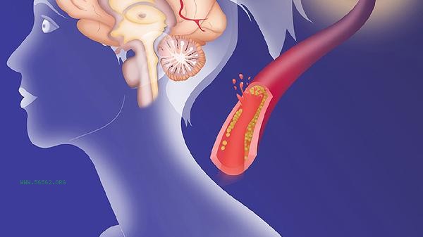
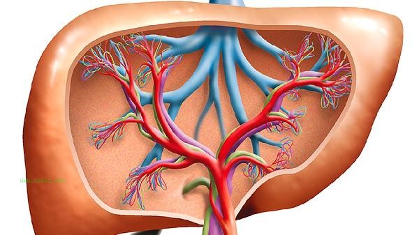
RI>0.75 indicates an increase in peripheral resistance, commonly seen in renal artery stenosis, cirrhosis, portal hypertension, or transplant organ rejection; A decrease in RI<0.55 may be seen in arteriovenous fistulas, tumors with abundant blood supply, or in a vasodilated state. It is necessary to make a comprehensive judgment by combining the pulsatility index PI and the systolic/diastolic ratio S/D.
4. Control of influencing factors:
Patient position, respiratory status, and probe pressure can all affect the measurement results. Before the examination, it is necessary to rest calmly for 10 minutes and avoid holding your breath or vigorous exercise. Vascular calcification, edema at the examination site, and other factors can interfere with signal acquisition. If necessary, use energy Doppler or contrast-enhanced ultrasound to assist in evaluation.
5. Equipment parameter settings:
uses a 5-12MHz high-frequency linear array probe to detect superficial blood vessels, and a 2-5MHz convex array probe for deep blood vessels. Adjust the Doppler gain until the background noise disappears, set the wall filter to 50-100Hz to eliminate vascular wall motion artifacts, and dynamically adjust the velocity scale based on blood flow velocity.
Regular vascular examination is recommended once a year, which can be shortened to half a year for patients with hypertension and diabetes. Maintain a low salt diet on a daily basis, with sodium intake controlled within 5g per day; Regular aerobic exercise such as brisk walking and swimming, with 150 minutes of moderate intensity exercise per week, can improve vascular elasticity; Smoking cessation and alcohol restriction, nicotine can directly damage the endothelial function of blood vessels. When abnormal RI is detected, further examinations such as CTA and MRA should be conducted to avoid self interpretation of the results and delay in diagnosis and treatment.


Comments (0)
Leave a Comment
No comments yet
Be the first to share your thoughts!